Abstract
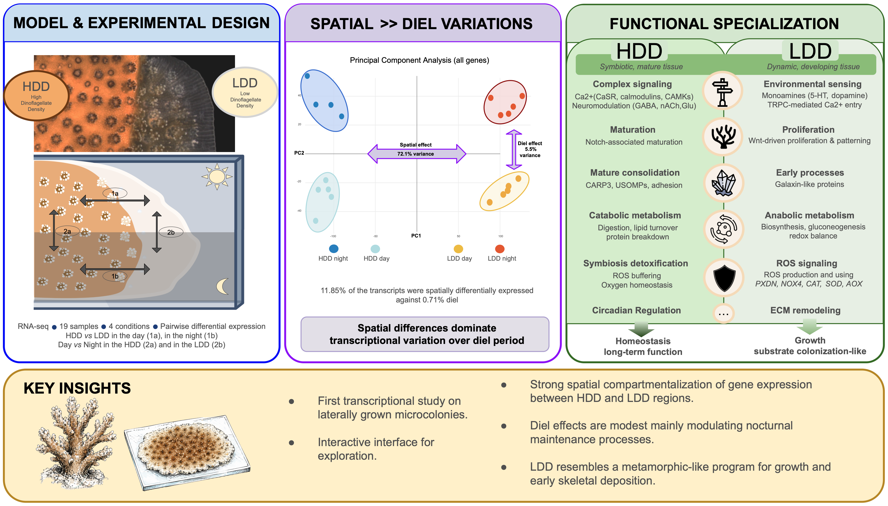
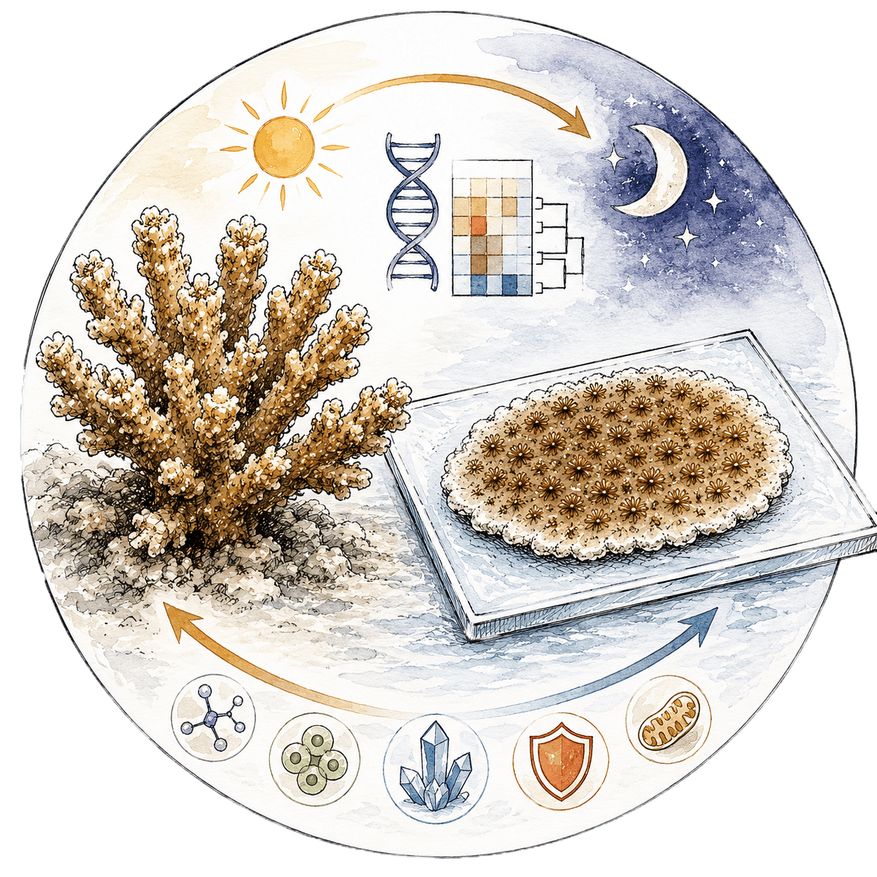
Corals grown laterally on glass slides have been used extensively as models to investigate aspects of coral cell biology, particularly coral biomineralization (Venn et al. 2024). Here we characterize transcriptomic variation between distinct tissue areas: low symbiotic density (LDD) areas at the transparent growing edge of the microcolony where the skeleton is in its earliest steps of development; and high dinoflagellate density (HDD) areas closer to the center of the microcolony where tissue is darkly pigmented and where the skeleton is mature. We investigated gene expression profiles from these areas during the day and at night to assess how light and tissue type interact to shape coral physiology. Transcriptomics analyses revealed strong spatial differentiation between LDD and HDD tissues, whereas diel effects were limited. While both regions share similar processes, they are characterized with specific actors. These findings suggest that growing-edge tissues transiently recapitulate developmental programs involved in substrate colonization, providing new insight into how spatially localized processes contribute to coral growth and skeletal formation.
In this tool, one can explore the HDD and LDD
Differentially
Expressed
Genes (
DEGs) during the day and the night but also comparing diel profiling of each regions.
This is accessible in
Explore.
A supplementary representation is available to
Discover genes we could not annotate in these four comparisons, the unknown DEGs... Providing an exciting opportunity to discover more!
 Coral Expression Atlas